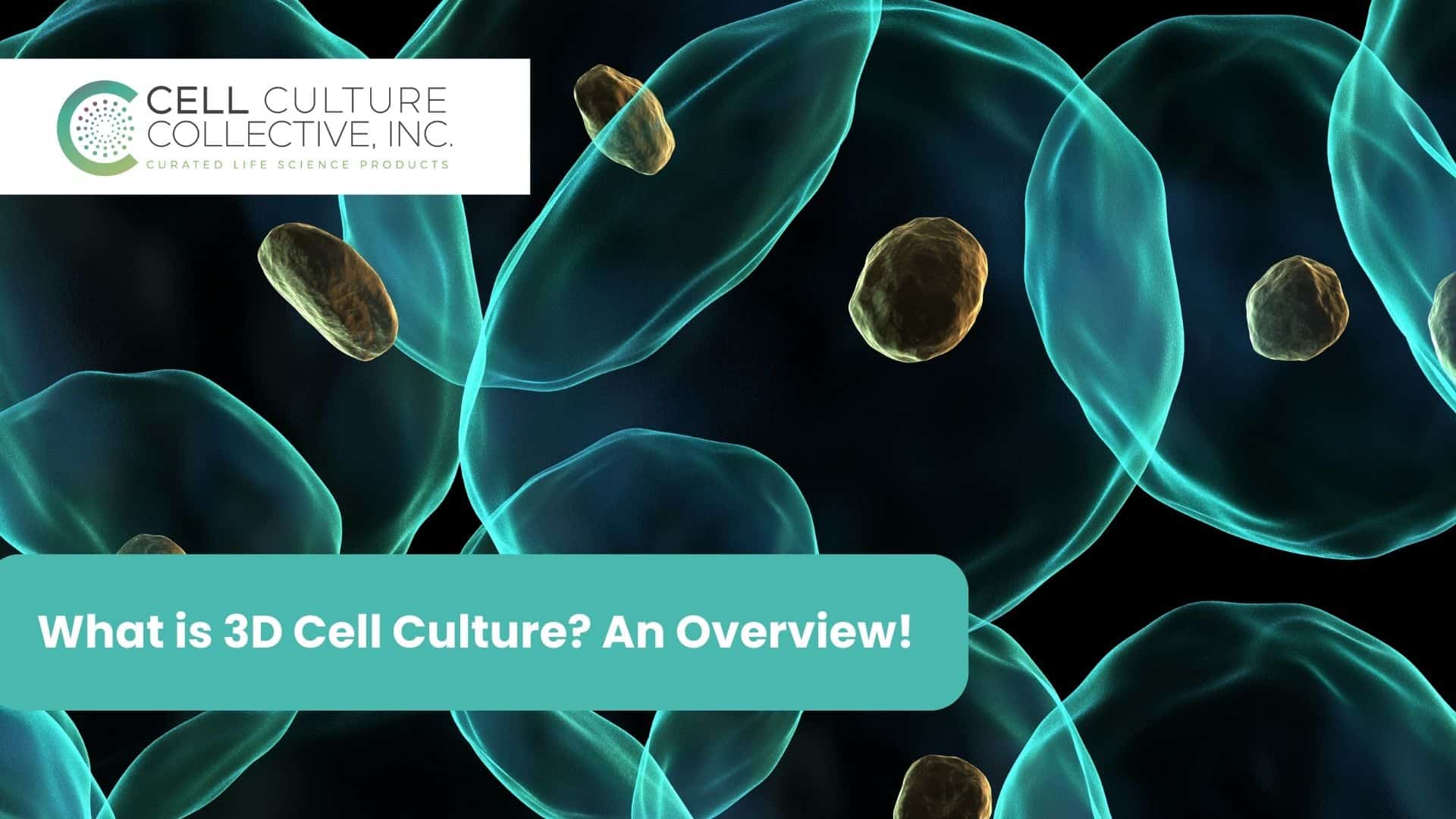
Imagine being able to replicate how cells behave inside the human body, but in a lab. That’s exactly what 3D cell culture is doing for researchers today. Unlike traditional 2D cultures, where cells are forced to grow flat, 3D cell culture allows them to interact in a way that closely mimics the body’s natural environment. And that’s a game-changer.
You see, the problem with 2D cultures is that they just don’t tell the full story. Cells in the body aren’t static. They are in constant motion, communicating, growing, and interacting with their surroundings in all directions. In fact, the 3D cell culture market is expected to skyrocket to $3.48 billion by 2028. Why? Because it’s helping researchers get much closer to real-life conditions, especially when it comes to drug discovery and personalized medicine.
For example, if you’re developing a cancer treatment, you need to understand how tumor cells behave in a 3D structure, not just on a flat plate. That’s where 3D cell culture steps in, offering more accurate results and helping to close the gap between what happens in the lab and what actually happens in the body.
It’s no wonder this approach is reshaping how we look at disease models and therapeutic testing. And, as the field continues to evolve, the possibilities for 3D cell culture seem almost endless.
What is 3D Cell Culture?
Think of 3D cell culture as giving cells the freedom to live and grow the way they would inside a living body. Traditional 2D cell culture methods have been around for decades, where cells are spread out flat on a plate, like they’re living on a sheet of glass. The problem? That flat world doesn’t quite capture the complexity of how cells really behave in our bodies. They miss out on the crucial interactions that happen in all directions: up, down, and sideways.
That’s where 3D cell culture comes in, offering cells a more life-like environment. In this setup, cells can communicate with their surroundings in all three dimensions, just as they do in the body. This more natural interaction leads to results that are closer to real-life conditions, making 3D cell culture a huge leap forward for researchers studying everything from drug responses to disease progression.
But not all 3D cultures are the same. Cells can grow in two major ways: scaffold-based and scaffold-free systems.
- Scaffold-based models act like a framework, giving cells a structure to cling to as they grow in every direction. Think of it as building a house with walls that guide the shape. Popular scaffolds include hydrogels, which provide a soft, water-rich environment much like human tissue, and inert matrices, which offer a solid surface for cells to grow into.
- On the other hand, scaffold-free models give cells the freedom to form their own structures. Without any external support, cells self-assemble into spheroids or clusters, mimicking the way tissue might grow in the body. Methods like low-adhesion plates and hanging drop cultures are perfect examples of how cells can grow without needing a scaffold to lean on.
In both cases, the goal is the same: to provide a more realistic environment for cells to behave as they would in their natural habitat, allowing scientists to study processes that 2D cultures simply can’t replicate.
Popular 3D Cell Culture Techniques
| Technique | Description | Applications |
| Scaffold-Based Techniques | ||
| Hydrogels | Water-rich polymer networks that mimic soft tissue environments. Can be derived from natural or synthetic sources like collagen or QGel® Matrix. | Tissue engineering, drug delivery, cancer research |
| Inert Matrices | Solid, porous structures like polystyrene membranes that provide a rigid environment for cell growth. | Long-term cell culture, stem cell research, differentiation studies |
| 3D Bioprinting | A cutting-edge method that uses bio-inks to print cells into complex 3D structures, enabling the construction of tissues layer by layer. | Tissue engineering, organ modeling, regenerative medicine |
| Scaffold-Free Techniques | ||
| Spheroid Formation | Cells naturally assemble into spherical clusters, replicating the in vivo tumor microenvironment. Achieved through low-adhesion plates or similar methods. | Tumor biology, drug testing, toxicology studies |
| Organoids | Miniature, simplified versions of organs that self-organize from stem cells, offering a more complex model than spheroids. | Disease modeling, personalized medicine, developmental biology |
| Hanging Drop Method | Uses gravity to form spheroids by placing cells in a suspended droplet, allowing for controlled aggregation and spheroid formation. | Precision spheroid formation, cancer research, drug discovery |
Advantages of 3D Cell Culture Over 2D Culture
When it comes to studying cells, the difference between 2D and 3D cultures is like night and day. In a traditional 2D environment, cells are stretched out and confined to a flat surface, which can dramatically affect how they behave. Think of it like asking someone to live their whole life lying down, they might survive, but they wouldn’t act the same way as if they were free to move around.
With 3D cell culture, cells get to live more naturally, interacting in all three dimensions: up, down, and sideways. Just like they would inside your body. This seemingly small change has a huge impact on research. Here’s why:
- More Realistic Cell Behavior
In a 3D environment, cells behave more like they do in real life. They form structures that resemble tissue, communicate with each other more effectively, and even react to drugs more predictably. For researchers, this means better, more accurate results, especially when testing new therapies or studying complex diseases. - Improved Drug Testing
Drug discovery relies heavily on how well a cell culture can replicate real-life conditions. Since 3D cell cultures allow cells to interact with their surroundings in a more natural way, they provide more reliable data on how drugs will work in human tissues. This is a big win for pharmaceutical companies, as it helps them identify potential failures earlier in the process which saves time, money, and even lives. - Better Modeling of Diseases
Diseases like cancer don’t just grow in a flat layer. They develop into complex, three-dimensional masses. By using 3D cell cultures, researchers can create more accurate models of tumors or diseased tissues, which leads to better understanding and more targeted treatments. - Mimicking the In Vivo Environment
In vivo means “within the living.” One of the biggest challenges with 2D cultures is that they fail to recreate the complexity of a living organism. In contrast, 3D cultures can mimic tissue stiffness, cell-to-cell interactions, and other key factors, providing a closer approximation to human physiology. - Stem Cell Research
When it comes to stem cell research, 3D cultures are particularly valuable. They help researchers better understand how stem cells differentiate and grow, which has major implications for regenerative medicine and the development of new treatments for various conditions.
In short, 3D cell culture isn’t just an upgrade, it’s a transformation in how we study cells. By moving away from flat, restrictive environments, scientists can now explore cell behavior in ways that bring them closer to understanding the complex, three-dimensional reality of life inside the human body.
Applications of 3D Cell Culture
The beauty of 3D cell culture lies in its versatility. It’s not just a tool for one specific field, it’s revolutionizing research across a range of disciplines. By offering a more accurate and life-like environment for cells to grow, 3D cell culture is helping scientists make breakthroughs that were once out of reach. Here are some of the key areas where 3D cell culture is making its mark:
- Drug Discovery and Development
The journey of creating a new drug is long, complex, and expensive, often with disappointing results. That’s where 3D cell culture comes in, offering a more accurate testing ground for new therapies. By mimicking the way cells behave inside the human body, 3D models can better predict how a drug will work in real tissues. This allows pharmaceutical companies to screen drugs faster and with greater precision, reducing the risk of failure in later stages of development.
- Cancer Research
Cancer is one of the most complex diseases to study because tumors grow in three dimensions, not just as flat layers of cells. 3D cancer models offer a more realistic way to study tumor behavior, including how cancer cells invade surrounding tissue and respond to treatments. This has massive implications for developing more effective cancer therapies, as it allows researchers to study tumors in conditions that are closer to those inside the human body. - Stem Cell Research and Regenerative Medicine
Stem cells have the unique ability to transform into many different types of cells, but studying them in 2D cultures often limits their potential. 3D cell culture opens up new possibilities in stem cell research, allowing scientists to explore how these cells grow, differentiate, and form tissues more effectively. This is particularly important in the field of regenerative medicine, where the goal is to replace or repair damaged tissues. - Personalized Medicine
Imagine being able to test a treatment on a miniature version of your own tissue before ever receiving it. With 3D organoid models, this is becoming a reality. By growing organoids from a patient’s stem cells, doctors can test how different therapies might work on a personalized level. This is a major leap forward in personalized medicine, offering tailored treatments that are specific to the individual. - Gene and Protein Expression Studies
In 2D cultures, gene and protein expression often don’t mirror what happens inside a living organism. However, in 3D cultures, cells experience conditions that are much closer to in vivo environments, leading to more accurate insights into how genes and proteins behave. This is crucial for understanding complex diseases and for developing treatments that target specific genetic pathways. - Toxicology and Safety Testing
Before any drug or chemical is approved for human use, it must undergo rigorous safety testing. 3D cell culture provides a better platform for toxicology studies, as it replicates how cells in human tissues would react to potential toxins. This reduces reliance on animal testing and offers more human-relevant data, leading to safer products.
In each of these areas, 3D cell culture is transforming how we study, understand, and treat diseases. By moving beyond flat cell layers, we’re gaining a deeper, more realistic view of how cells behave, offering new insights and possibilities that are shaping the future of medicine.
Conclusion
The shift from 2D to 3D cell culture is more than just a technical upgrade. Tt’s a complete transformation in how we understand biology. By giving cells the space to grow and interact as they would in the human body, 3D cell culture offers a level of realism that opens doors to groundbreaking discoveries. Whether it’s in drug development, cancer research, or personalized medicine, this approach is paving the way for more accurate studies, faster drug approvals, and even patient-specific treatments.
As research continues to evolve, the possibilities seem endless. We’re seeing advancements like organoids and bioprinting that are pushing the boundaries of what we thought was possible in cell biology. And with the global market for 3D cell culture expected to grow exponentially, it’s clear that this is more than just a trend, it’s the future of medical research.
In the end, 3D cell culture isn’t just helping us answer the questions we already have. It’s allowing us to ask new ones, leading to discoveries that could change the way we approach health and disease for generations to come.

