Cell culture technique is more than just a laboratory experiment; it’s a foundation of modern biological research. From studying basic cell behavior to pioneering new treatments, mastering cell culture techniques opens doors to discoveries that drive science forward. Whether you’re culturing mammalian cells for disease research, maintaining strict aseptic conditions, or experimenting with advanced 3D models, each step plays a crucial role in ensuring that your research reflects true-to-life results.
In this guide, we’ll explore the essentials of cell culture techniques, moving from foundational practices to advanced applications that mimic complex biological environments. Whether you’re here to sharpen your skills or dive into new methods, this journey into cell culture will cover everything you need to know from keeping your cultures contaminant-free to scaling up for more sophisticated, in vivo-like studies.
Basics of Cell Culture Techniques
Introduction to Cell Culture
At its core, cell culture is the process of growing cells in a controlled environment outside their natural setting. This technique allows scientists to study cell behavior, conduct drug testing, and model diseases in a lab setting that mimics real-life conditions. Whether researchers are investigating cell growth, gene expression, or cellular interactions, cell culture is foundational to understanding biological processes at the cellular level.
Key Components
Creating an effective cell culture setup requires specific tools and environmental controls to ensure cell health and viability. Here are the essentials:
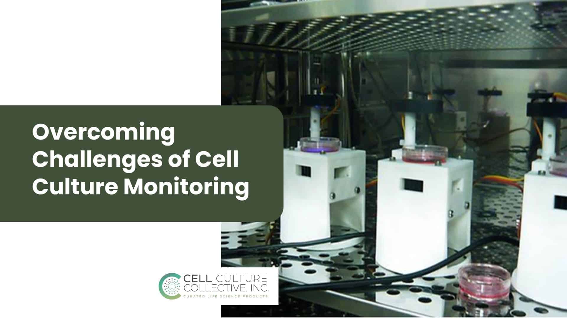
- Incubators: Maintain ideal temperature (usually 37°C for mammalian cells) and humidity levels to mimic the body’s internal conditions.
- Growth Media: Nutrient-rich solutions like DMEM or RPMI, customized to support cell growth by providing necessary vitamins, amino acids, and other factors.
- Culture Vessels: Various types, including petri dishes, flasks, and multi-well plates, depending on the type of cells and experiment requirements.
- Environmental Controls: In addition to temperature, CO₂ levels are controlled (typically at 5%) to stabilize the pH, keeping the culture environment as close to in vivo conditions as possible.
Each component is critical for establishing a stable culture environment where cells can thrive and yield reliable results.
Cell Types
In cell culture, different cell types are used depending on the research goals. Among these, mammalian cells are some of the most commonly cultured due to their relevance in human biology. Mammalian cell culture techniques focus on keeping cells alive and functional, providing insights into cellular responses, disease mechanisms, and therapeutic testing. Researchers may work with:
- Primary Cells: Cells taken directly from a living organism, which maintain many of their natural characteristics but have a limited lifespan.
- Immortalized Cell Lines: Cells that have been genetically altered or naturally evolved to grow indefinitely, ideal for experiments requiring repeated trials.
- Stem Cells: Versatile cells that can differentiate into various cell types, often used in regenerative medicine and developmental biology studies.
Each cell type has unique needs, but understanding their characteristics is key to setting up experiments that yield meaningful insights.
Aseptic Techniques in Cell Culture
Importance of Aseptic Technique
In cell culture, maintaining a sterile environment is critical. Aseptic technique prevents contamination from microorganisms and cross-contamination between different cell lines, which can compromise the health of the culture and skew results. Even a small lapse in aseptic practices can introduce bacteria, fungi, or other pathogens, leading to inaccurate data or even culture loss. Practicing good aseptic technique safeguards the integrity of experiments, ensuring that cells remain healthy and uncontaminated.
Steps in Aseptic Technique
Aseptic technique involves a series of careful steps designed to minimize contamination:
- Sterilization
- Sterilize all equipment and media before use. This includes autoclaving reusable tools, using sterile disposable pipettes, and filtering media to eliminate any potential contaminants.
- Laminar Flow Hood
- Perform all cell culture work in a laminar flow hood, which creates a sterile airflow to protect the culture. Disinfect the hood surface before and after each use with 70% ethanol to ensure it remains sterile.
- Personal Hygiene and Preparation
- Wash hands thoroughly, wear gloves, and avoid touching any surfaces that aren’t sterile. Gloves and other protective equipment should be sanitized with ethanol before entering the culture area.
- Proper Handling Techniques
- Work with open containers as briefly as possible to limit exposure to air. Avoid reaching over open containers, and keep culture vessels closed when not in use.
By following these steps, researchers maintain a contaminant-free environment, critical for the health and reliability of cell cultures.
Common Contaminants
Despite best efforts, contaminants can sometimes enter cultures. Here are some of the most common types and tips for prevention:
- Bacterial Contamination: The most frequent type, often visible as a cloudy medium or changes in pH. Prevented by stringent aseptic technique and regularly cleaning surfaces and equipment.
- Fungal Contamination: Fungi appear as filamentous growths in the culture and can be introduced from air or improperly sterilized items. Use of antifungal agents in media can help minimize this risk.
- Viral Contamination: Less common but challenging to detect and eliminate, often requiring specific viral screening protocols to identify. This risk is minimized by sourcing cells from reputable providers.
- Cross-Contamination: When cells from one culture mix with another, often due to inadequate sterilization of shared equipment. Strictly following aseptic procedures and using separate, clearly labeled equipment can help prevent this.
Each step and preventive measure in aseptic technique helps researchers keep their cell cultures clean and dependable, which is crucial for accurate and reproducible results.
Core Mammalian Cell Culture Techniques
Passaging and Subculturing
To maintain healthy mammalian cell lines, passaging or subculturing is essential. This process involves splitting cells when they reach a certain density to prevent overcrowding and keep the culture in a growth-friendly state. Here’s how it’s done:
- Cell Detachment: Cells are detached from the culture surface using trypsin, an enzyme that breaks down cell adhesion.
- Cell Counting: Once detached, cells are counted to determine the appropriate density for the next culture.
- Re-Seeding: Cells are then transferred to new culture vessels at a lower density to allow room for further growth. Regular passaging helps prevent the cells from reaching senescence too soon and ensures their continued proliferation.
Seeding and Cell Density
Achieving the right cell density during seeding is critical for optimal growth and health. If cells are seeded too densely, they compete for resources, leading to stress and early senescence. If seeded too sparsely, they may not thrive due to a lack of cell-cell interactions that promote healthy growth. Key considerations include:
- Optimal Density: Seeding densities vary by cell type, but maintaining the right density encourages balanced growth.
- Monitoring Confluence: Regularly monitor the culture for confluence (the coverage of the culture surface), aiming for around 70-80% confluence before passaging.
Setting up the correct seeding density ensures that cells grow at an ideal rate, maintaining viability and productivity in the culture.
Media Changes and Monitoring
For cells to remain in peak condition, regular media changes are necessary to replenish nutrients and remove waste products. Over time, cells consume nutrients and release metabolic waste, which can affect cell health if not managed properly. The media change process includes:
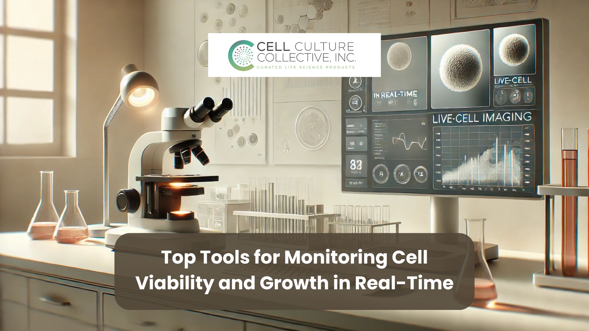
- Observing Growth Patterns: Before changing the media, observe the culture under a microscope for any signs of contamination or unusual growth behavior.
- Changing Media: Remove the spent media carefully and replace it with fresh, pre-warmed media to prevent temperature shock.
- Routine Monitoring: Keep an eye on cell morphology, growth rate, and color changes in the media, which may indicate pH shifts or contamination.
These techniques, passaging, seeding at optimal density, and routine media changes, form the foundation of mammalian cell culture, enabling researchers to maintain cell health and produce reliable, consistent results.
Advanced Cell Culture Techniques
3D Cell Culture and Organoids
Traditional cell culture often involves growing cells in a flat, 2D environment. However, 3D cell culture techniques provide a more in vivo-like setting, allowing cells to grow in three dimensions. This approach enables cells to interact more naturally with their surroundings, simulating the complex architecture of tissues within the body.
- Organoids: One of the most innovative applications of 3D culture, organoids are miniature, self-organizing structures derived from stem cells that mimic the functions of actual organs. These mini-organs allow researchers to study organ-specific processes, test drugs, and explore disease mechanisms with remarkable accuracy.
- Advantages of 3D Cultures: By offering a closer resemblance to real tissue environments, 3D cultures improve the relevance of research findings, making them ideal for cancer studies, regenerative medicine, and drug testing.
Co-Culture Systems
Co-culture systems involve cultivating multiple cell types within the same environment, allowing scientists to study cell-cell interactions that are crucial in many biological processes. This technique is particularly useful for understanding complex systems like the immune response, where multiple cell types work in concert.
- Applications: Co-cultures are widely used in cancer research to study tumor-stromal interactions, as well as in neuroscience to examine neuron-glia interactions. They are also essential in studying tissue engineering and immune system functions.
- Enhanced Insights: By recreating natural cell interactions, co-cultures offer insights that single-cell-type cultures cannot provide, helping researchers better understand disease mechanisms and cellular responses in a more holistic way.
Microfluidics and Organs-on-Chips
The most sophisticated of advanced cell culture techniques, microfluidics and organs-on-chips bring new levels of complexity and precision to in vitro models. These platforms use channels and tiny chambers to introduce fluid flow, mimicking blood circulation and creating conditions that closely resemble in vivo environments.
- Organs-on-Chips: These are microfluidic devices containing living cells that model specific organ functions. For instance, a lung-on-a-chip might simulate the breathing motion and blood flow of a human lung, offering a powerful model for studying respiratory diseases or testing drug responses.
- Benefits of Fluid Dynamics: Integrating fluid flow in cell culture provides insights into how cells respond to shear stress, nutrient gradients, and other mechanical forces, enabling more accurate disease modeling, especially for cardiovascular and respiratory studies.
Applications of Cell Culture Techniques
Drug Discovery and Toxicology
Cell culture is fundamental in drug discovery and toxicology, providing a controlled environment to screen and test new drugs. Cultured cells allow researchers to evaluate a compound’s effectiveness and detect any toxic side effects early in the development process. By using cell cultures, scientists can observe how a drug interacts with cells, assess cellular responses, and determine safe dosage levels. This preclinical testing phase helps identify promising compounds before they move to animal or human trials, saving time and resources.
Cancer Research
In cancer research, cell culture techniques are crucial for studying tumor biology and testing potential treatments. By culturing cancer cells, researchers can investigate how tumors grow, mutate, and respond to various therapies, gaining insights into cellular mechanisms like proliferation, apoptosis (programmed cell death), and resistance. Cell culture enables scientists to test chemotherapy drugs, targeted therapies, and immunotherapies on cancer cells directly, helping to tailor treatments and understand their effects on specific types of cancer. This approach accelerates the development of personalized medicine, aiming to improve treatment outcomes for cancer patients.
Regenerative Medicine and Tissue Engineering
Cell culture techniques are at the forefront of regenerative medicine and tissue engineering. In this field, cells are used to create tissues and even organs that can replace damaged or diseased parts of the body. By cultivating stem cells and other cell types, scientists can encourage these cells to differentiate and form functional tissue structures, such as skin, cartilage, or even liver tissue. These cultured tissues serve as models for studying regeneration and hold promise for treating injuries, degenerative diseases, and organ failure. Ultimately, cell culture is paving the way for breakthroughs in creating lab-grown tissues that could be transplanted into patients, potentially transforming medical treatments.
Conclusion
Mastering cell culture techniques is a journey that bridges foundational science with cutting-edge research, opening doors to breakthroughs in drug discovery, cancer treatment, and regenerative medicine. From the basics of maintaining a sterile environment to the complexity of advanced 3D cultures and organs-on-chips, each technique has a unique role in advancing our understanding of human biology.
As cell culture techniques continue to evolve, so do the possibilities for creating more accurate, in vivo-like models that deepen our insights and enhance the relevance of research. Whether you’re exploring cellular responses to new treatments or engineering tissues for regenerative therapies, mastering these methods is key to making meaningful strides in science and medicine.


