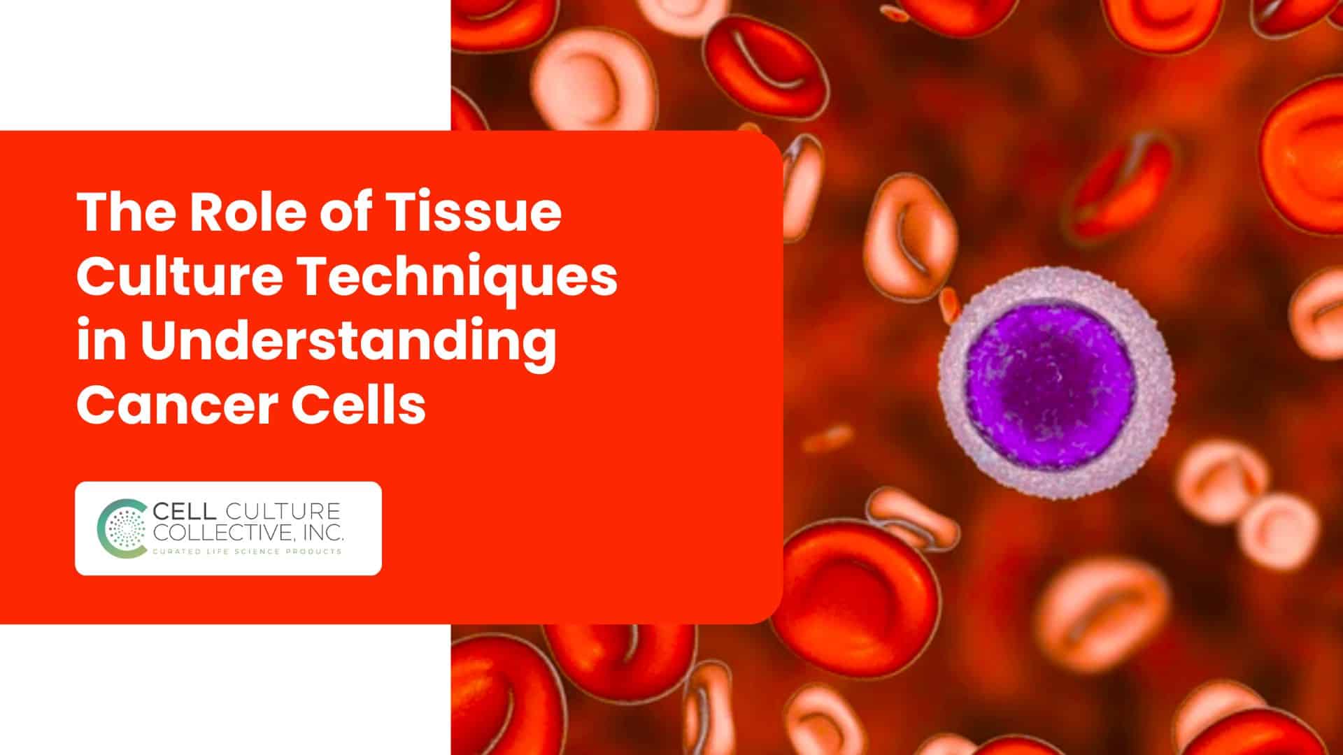
In the battle against cancer, understanding the very cells that drive the disease is a crucial step. Tissue culture techniques have become indispensable tools in cancer research, allowing scientists to grow and study cancer cells in controlled lab environments. By culturing cancer cells, researchers can observe their behaviors up close including how they grow, adapt, and sometimes resist treatments, all without the constraints of the human body.
From traditional 2D cultures to advanced 3D models that better mimic real tumors, these techniques provide insights that guide everything from basic research to cutting-edge therapies. In this guide, we’ll explore how tissue culture techniques have transformed our approach to studying cancer, revealing the hidden dynamics of tumor growth, drug resistance, and the interactions that shape the tumor microenvironment.
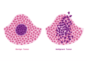
Source: National Cancer Foundation
What is Cancer Cell Culture?
Cancer cell culture is the process of growing cancer cells outside their natural environment, typically in a lab setting, to study their behavior, genetic changes, and responses to treatments. By cultivating these cells, researchers can simulate conditions that allow for closer examination of cancer cell dynamics, gaining insights that might be impossible to observe directly in the human body.
Importance of Tissue Culture in Cancer Studies
Culturing cancer cells provides a controlled environment where scientists can investigate the complexities of cancer at a cellular level. This technique allows researchers to:
- Observe growth patterns and mutations.
- Study interactions between cancer cells and treatments.
- Test drug efficacy and resistance, ultimately contributing to more targeted therapies.
Through tissue culture, scientists can manipulate variables with precision, making it an invaluable tool for developing effective cancer treatments.
Types of Cancer Cell Cultures
Cancer research relies on two primary types of cell culture methods:
- 2D Cultures: In these traditional methods, cancer cells are grown on a flat surface, like a petri dish or flask, where they form a single layer. Although widely used, 2D cultures often lack the complexity of the natural tumor environment.
- 3D Cultures: 3D culture techniques, including spheroids and organoids, allow cancer cells to grow in all directions, forming structures that more closely resemble actual tumors. This method enables more accurate studies of cell behavior, treatment responses, and the tumor microenvironment, making it particularly valuable for advanced cancer research.
The Process of Culturing Cancer Cells
Sample Collection and Preparation
The first step in culturing cancer cells is obtaining samples, which can come from solid tumors (through biopsies) or fluid samples (like blood or pleural effusions). These samples are carefully transported and processed in a sterile environment to ensure viability. Once in the lab, the tissue undergoes mechanical disaggregation (using scalpels to cut into smaller fragments) and enzymatic digestion (using enzymes like collagenase or trypsin to break down extracellular matrix components). This dual approach helps release individual cancer cells, which are then placed in a culture medium to support their growth.
Culturing Techniques
2D Culture Methods
2D cell cultures are the traditional method in cancer research, where cells grow on a flat, two-dimensional surface like a petri dish or flask. Cells in 2D cultures form a monolayer and are directly exposed to the nutrients in the culture medium, making them easy to observe and manipulate. These methods are commonly used for:
- Initial drug screenings: Testing how cancer cells respond to new compounds.
- Basic studies: Examining fundamental cell behaviors such as growth rates and gene expression.
However, the limitations of 2D cultures lie in their inability to mimic the complex architecture and interactions within a real tumor, often leading to differences in how cells respond in vivo versus in vitro.
3D Culture Methods
3D cell cultures have become increasingly popular due to their ability to mimic in vivo conditions more accurately. These methods allow cancer cells to grow in three dimensions, forming structures similar to actual tumors. There are several 3D techniques:
- Spheroids: Cancer cells self-assemble into spherical structures that can mimic the oxygen and nutrient gradients found in tumors.
- Organoids: Stem cell-derived or cancer cell-derived structures that resemble specific organs, allowing researchers to study how tumors might develop in particular tissues.
- Scaffold-Based Systems: Cancer cells grow on synthetic or natural scaffolds, providing structural support that allows them to organize and behave like they would in the body.
3D culture methods are especially useful for studying cancer cell interactions, drug resistance, and treatment efficacy in a more realistic context, offering insights that 2D cultures cannot provide.
Aseptic Techniques
Maintaining sterility is essential in cancer cell culture to prevent contamination and ensure reliable results. Aseptic techniques include sterilizing all equipment, working in a laminar flow hood, and wearing gloves and other protective equipment to reduce contamination risks. By following strict aseptic protocols, researchers can minimize bacterial, fungal, and viral contamination, which could compromise the integrity of cancer cell studies and lead to false results.
Key Insights Gained from Cancer Cell Culture
Understanding Tumor Behavior
- Provides insights into cancer cell proliferation (how quickly cells divide and grow).
- Studies invasion mechanisms and how cancer cells penetrate nearby tissues.
- Examines metastasis, the process by which cancer cells spread to distant organs.
- Allows for detailed observation of cellular changes over time, offering a better understanding of tumor progression.
Drug Screening and Resistance Studies
- Enables drug efficacy testing on cultured cancer cells before advancing to animal or human trials.
- Helps identify drug resistance mechanisms by observing how cancer cells adapt to treatments.
- Supports the development of combination therapies by testing different drugs together in controlled settings.
- Increases treatment effectiveness by tailoring drug responses to specific cancer cell types.
Tumor Microenvironment Studies
- 3D cultures mimic the complex structure of actual tumors, providing a realistic environment for study.
- Allows researchers to observe cell-cell interactions and cell-matrix interactions, crucial for tumor development.
- Facilitates studies on immune cell interactions with cancer cells, essential for immunotherapy research.
- Enhances understanding of how the microenvironment influences cancer growth, drug response, and cell behavior.
Advanced Techniques Enhancing Cancer Research
In cancer research, advanced tissue culture techniques have expanded the possibilities of what scientists can observe and understand about tumors. From dynamic models that mimic blood flow to storing cells for personalized treatment testing, these cutting-edge methods offer tools to study cancer with remarkable accuracy.
Organs-on-Chips and Microfluidics
- Organs-on-Chips use microfluidic devices to create small, functional models of human organs, integrating live cancer cells in a controlled flow of nutrients and oxygen.
- By simulating blood flow, microfluidic devices replicate the tumor’s dynamic environment, allowing researchers to observe cancer cell behavior under conditions similar to those in the body.
- Applications: These devices enable studies on drug delivery, metastasis, and tumor cell responses to changes in their environment, which are critical for developing targeted therapies.
Co-Cultures with Immune Cells
- Co-culture systems bring together cancer cells and immune cells, enabling researchers to study interactions that are crucial to understanding tumor immunity.
- Importance: Co-cultures reveal how immune cells recognize and attack cancer cells, and they play a major role in developing immunotherapies that enhance the body’s natural defenses.
- Applications: By examining cancer-immune cell dynamics, scientists can test how tumors evade immune responses and how to improve treatment strategies that harness the immune system.
Cryopreservation and Biobanking
- Cryopreservation involves freezing cancer cells at ultra-low temperatures, allowing them to be stored long-term without losing viability.
- Biobanking: This approach enables the storage of cancer cells from various patients, creating a repository for future research and testing.
- Applications: Cryopreserved cells are used in personalized medicine for tailored drug testing and in studies requiring consistent cell populations over time.
Conclusion
The journey of understanding cancer is one of relentless innovation and precision, and tissue culture techniques lie at the very heart of this quest. From traditional methods that laid the groundwork to cutting-edge 3D cultures and organs-on-chips that bring us as close as possible to real tumors, each technique opens new doors in cancer research. These methods don’t just allow us to study cells in isolation; they let us explore the complexities of canceri all within a controlled environment that reveals its secrets, one culture at a time.
As we continue to push the boundaries with advanced co-cultures, personalized biobanks, and simulated microenvironments, the potential for breakthroughs in cancer therapy grows exponentially. Every cultured cell, every preserved sample, every microfluidic chip adds to a growing arsenal against one of humanity’s most formidable foes. And in this relentless pursuit, we’re not just aiming for treatments but for truly transformative solutions that will rewrite what’s possible in cancer care. The future of cancer research is bright, dynamic, and evolving faster than ever, and tissue culture techniques are lighting the way.

